 Mixing
Saltatory conduction

Action potential propagation in myelinated neurons is faster than in unmyelinated neurons because of saltatory conduction.
| Saltatory conduction occurs only on myelinated axons. |
|---|
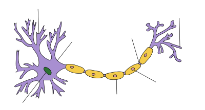 Dendrite Soma Axon Nucleus Node of Ranvier Axon terminal Schwann cell Myelin sheath |

Propagation of action potential along myelinated nerve fiber
Saltatory conduction (from the Latin saltare, to hop or leap) is the propagation of action potentials along myelinated axons from one node of Ranvier to the next node, increasing the conduction velocity of action potentials. The uninsulated nodes of Ranvier are the only places along the axon where ions are exchanged across the axon membrane, regenerating the action potential between regions of the axon that are insulated by myelin, unlike electrical conduction in a simple circuit.
Contents
1 Mechanism
2 Energy efficiency
3 Distribution
4 See also
5 References
6 Further reading
7 External links
Mechanism
Myelinated axons only allow action potentials to occur at the unmyelinated nodes of Ranvier that occur between the myelinated internodes. It is by this restriction that saltatory conduction propagates an action potential along the axon of a neuron at rates significantly higher than would be possible without the myelination of the axon (150 m/s compared to 0.5 to 10 m/s).[1] As sodium rushes into the node it creates an electrical force which pushes on the ions already inside the axon. This rapid conduction of electrical signal reaches the next node and creates another action potential, thus refreshing the signal. In this manner, saltatory conduction allows electrical nerve signals to be propagated long distances at high rates without any degradation of the signal. Although the action potential appears to jump along the axon, this phenomenon is actually just the rapid, almost instantaneous, conduction of the signal inside the myelinated portion of the axon.
Energy efficiency
In addition to increasing the speed of the nerve impulse, the myelin sheath helps in reducing energy expenditure over the axon membrane as a whole, because the amount of sodium and potassium ions that need to be pumped to bring the concentrations back to the resting state following each action potential is decreased.[2]
Distribution
Saltatory conduction occurs widely in the myelinated nerve fibers of vertebrates, but was later discovered in a pair of medial myelinated giant fibers of Fenneropenaeus chinensis and Marsupenaeus japonicus shrimp,[3][4][5] as well as in a median giant fiber of an earthworm.[6] Saltatory conduction has also been found in the small- and medium-sized myelinated fibers of Penaeus shrimp.[7]
See also
- Bioelectrochemistry
- Cable theory
- Electrophysiology
- Ephaptic coupling
- GHK current equation
- Goldman equation
- Hindmarsh–Rose model
- Hodgkin–Huxley model
- Neurotransmission
- Patch clamp
- Quantitative models of the action potential
References
^ Purves D, Augustine GJ, Fitzpatrick D (2001). "Increased Conduction Velocity as a Result of Myelination". Neuroscience (2nd ed.). Sunderland (MA): Sinauer Associates..mw-parser-output cite.citation{font-style:inherit}.mw-parser-output q{quotes:"""""""'""'"}.mw-parser-output code.cs1-code{color:inherit;background:inherit;border:inherit;padding:inherit}.mw-parser-output .cs1-lock-free a{background:url("//upload.wikimedia.org/wikipedia/commons/thumb/6/65/Lock-green.svg/9px-Lock-green.svg.png")no-repeat;background-position:right .1em center}.mw-parser-output .cs1-lock-limited a,.mw-parser-output .cs1-lock-registration a{background:url("//upload.wikimedia.org/wikipedia/commons/thumb/d/d6/Lock-gray-alt-2.svg/9px-Lock-gray-alt-2.svg.png")no-repeat;background-position:right .1em center}.mw-parser-output .cs1-lock-subscription a{background:url("//upload.wikimedia.org/wikipedia/commons/thumb/a/aa/Lock-red-alt-2.svg/9px-Lock-red-alt-2.svg.png")no-repeat;background-position:right .1em center}.mw-parser-output .cs1-subscription,.mw-parser-output .cs1-registration{color:#555}.mw-parser-output .cs1-subscription span,.mw-parser-output .cs1-registration span{border-bottom:1px dotted;cursor:help}.mw-parser-output .cs1-hidden-error{display:none;font-size:100%}.mw-parser-output .cs1-visible-error{font-size:100%}.mw-parser-output .cs1-subscription,.mw-parser-output .cs1-registration,.mw-parser-output .cs1-format{font-size:95%}.mw-parser-output .cs1-kern-left,.mw-parser-output .cs1-kern-wl-left{padding-left:0.2em}.mw-parser-output .cs1-kern-right,.mw-parser-output .cs1-kern-wl-right{padding-right:0.2em}
^ Tamarkin D. "Saltatory Conduction of APs". Archived from the original on 30 October 2014. Retrieved 6 May 2014.
^ Hsu K, Tan TP, Chen FS (August 1964). "On the excitation and saltatory conduction in the giant fiber of shrimp (Penaeus orientalis)". Proceedings of the 14th National Congress of the Chinese Association for Physiological Science: 7–15.
^ Hsu K, Tan TP, Chen FS (1975). "Saltatory conduction in the myelinated giant fiber of shrimp (Penaeus orientalis)". KexueTongbao. 20: 380–382.
^ Kusano K, LaVail MM (August 1971). "Impulse conduction in the shrimp medullated giant fiber with special reference to the structure of functionally excitable areas". The Journal of Comparative Neurology. 142 (4): 481–94. doi:10.1002/cne.901420406. PMID 5111883.
^ Günther J (August 1976). "Impulse conduction in the myelinated giant fibers of the earthworm. Structure and function of the dorsal nodes in the median giant fiber". The Journal of Comparative Neurology. 168 (4): 505–31. doi:10.1002/cne.901680405. PMID 939820.
^ Xu K, Terakawa S (1993). "Saltatory conduction and a novel type of excitable fenestra in shrimp myelinated nerve fibers". The Japanese Journal of Physiology. 43 Suppl 1: S285–93. PMID 8271510.
Further reading
.mw-parser-output .refbegin{font-size:90%;margin-bottom:0.5em}.mw-parser-output .refbegin-hanging-indents>ul{list-style-type:none;margin-left:0}.mw-parser-output .refbegin-hanging-indents>ul>li,.mw-parser-output .refbegin-hanging-indents>dl>dd{margin-left:0;padding-left:3.2em;text-indent:-3.2em;list-style:none}.mw-parser-output .refbegin-100{font-size:100%}
Saladin K. "Saltatory conduction". Biology Online.
Anatomy & physiology : the unity of form and function (6th ed.). McGraw-Hill. 2011. ISBN 978-0-07-768033-6.
External links
- Saltatory conduction - Scholarpedia
- cell biology - Why is saltatory conduction in myelinated axons faster than continuous conduction in unmyelinated axons?


Comments
Post a Comment