绿色荧光蛋白
| 绿色荧光蛋白 (Green fluorescent protein) | |||||||||
|---|---|---|---|---|---|---|---|---|---|
 | |||||||||
| 維多利亞多管水母綠色熒光蛋白的結構。[1] | |||||||||
| 鉴定 | |||||||||
| 标志 | Reginal | ||||||||
Pfam(蛋白家族查询站) | PF01353 | ||||||||
Pfam宗系 | CL0069 | ||||||||
InterPro(蛋白数据整合站) | IPR011584 | ||||||||
SCOP(蛋白结构分类数据站) | 1ema | ||||||||
| |||||||||

绿色荧光蛋白 (GFP)帶狀圖,來自 PDB 1EMA。

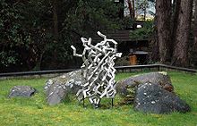
綠色熒光蛋白為基礎的水母鋼鐵雕塑(2006年)陳列在聖胡安島星期五港實驗室(華盛頓,美國),GFP的發現的地方。
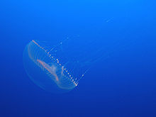
綠色螢光蛋白(Green fluorescent protein,簡稱GFP),是一个由约238个氨基酸组成的蛋白質,從藍光到紫外线都能使其激發,發出綠色螢光。[2][3]虽然许多其他海洋生物也有类似的绿色荧光蛋白,但傳統上,绿色荧光蛋白(GFP)指首先从維多利亞多管發光水母中分离的蛋白质。這種蛋白質最早是由下村脩等人在1962年在維多利亞多管發光水母中發現。這個發光的過程中還需要冷光蛋白質水母素的幫助,且這個冷光蛋白質與鈣離子(Ca2+)可產生交互作用。
在維多利亞多管發光水母中發現的野生型綠色螢光蛋白,395nm和475nm分別是最大和次大的激发波长,它的发射波長的峰點是在509nm,在可見光譜中處於綠光偏藍的位置。绿色荧光蛋白的荧光量子产率(QY)为0.79。而從海腎(sea pansy)所得的綠色螢光蛋白,僅在498nm有一個較高的激發峰點。
在細胞生物學與分子生物學中,綠色螢光蛋白(GFP)基因常用做報導基因(reporter gene)。[4],綠色螢光蛋白基因也可以轉殖到脊椎動物(例如:兔子)上進行表現,並拿來映證某種假設的實驗方法。通過基因工程,綠色螢光蛋白(GFP)基因能穩轉進不同物種的基因組,在後代中持續表達。現在,綠色螢光蛋白(GFP)基因已被导入并表达在许多物種,包括细菌,酵母和其他真菌,鱼(例如斑马鱼),植物,苍蝇,甚至人等的哺乳动物细胞。
2008年10月8日,日本科学家下村脩、美国科学家马丁·查尔菲和钱永健因为发现和改造绿色荧光蛋白而获得了当年的诺贝尔化学奖。[5][6]
目录
1 歷史
1.1 野生型GFP(wtGFP)
1.2 GFP衍生物
2 結構
3 应用
3.1 荧光显微镜
4 參見
5 參考資料
6 延伸閱讀
7 外部連結
歷史

維多利亞多管發光水母
野生型GFP(wtGFP)
在1960年代和1970年代,綠色螢光蛋白,連同分開發光蛋白水母素,首先從維多利亞多管發光水母被純化,及其屬性被下村修研究。[7]
GFP衍生物

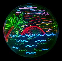
聖地亞哥海灘畫說明基因突變的多樣性,活細菌表達8種不同顏色的螢光蛋白。
由於對廣泛使用的潛力和研究人員不斷變化的需求,綠色螢光蛋白的許多不同的突變體已被改造設計。[8]
結構
野生型綠色熒光蛋白,最開始是 238 個氨基酸的肽鏈,約 25KDa。然後按一定規則,11 條β-摺疊在外周圍成圓柱狀的柵欄;圓柱中,α-螺旋把發色團固定在正幾乎中心處。發色圖被圍在中心,能避免偶極化的水分子、順磁化的氧分子或者順反異構作用與發色團,致使熒光猝滅。[3]
熒光是熒光蛋白最特別的特點,而其中的髮色團起着主要的作用。在 α-螺旋上的 65、66、67位氨基酸——絲氨酸、酪氨酸、甘氨酸經過環化、脫氫等作用後形成發色團。有意思的是,發色團形成過程是由外周柵欄上的殘基催化,底物只需要氧氣。[1]這暗示綠色熒光蛋白被廣泛用於不同物種的潛力:在不同物種中能獨立表達成有功能的蛋白,而不需要額外的因子。不過,現在依然在討論準確的過程。[9]
發色團上的共軛 π鍵能吸收激發光能量,在很短的時間後,以波長更長的發射光釋放能量,形成熒光。
应用
由於熒光蛋白能穩定在後代遺傳,並且能根據啓動子特異性地表達,在需要定量或其他實驗中慢慢取代了傳統的化學染料。更多地,熒光蛋白被改造成了不同的新工具,既提供了解決問題的新思路,也可能帶來更多有價值的新問題。
荧光显微镜
GFP和它的衍生物的可用性已经彻底重新定义荧光显微镜,以及它被用来在细胞生物学和其他生物学科的方式。[10]。其中,最令人興奮的就是用於超分辨顯微鏡成像。
參見
- pGLO
- 黃色熒光蛋白
參考資料
^ 1.01.1 Ormö M, Cubitt AB, Kallio K, Gross LA, Tsien RY, Remington SJ. Crystal structure of the Aequorea victoria green fluorescent protein. Science. September 1996, 273 (5280): 1392–5. PMID 8703075. doi:10.1126/science.273.5280.1392.
^ Prendergast FG, Mann KG; Mann. Chemical and physical properties of aequorin and the green fluorescent protein isolated from Aequorea forskålea. Biochemistry. 1978, 17 (17): 3448–53. PMID 28749. doi:10.1021/bi00610a004.
^ 3.03.1 Tsien RY. The green fluorescent protein (PDF). Annu Rev Biochem. 1998, 67: 509–44. PMID 9759496. doi:10.1146/annurev.biochem.67.1.509.
^ Phillips G. Green fluorescent protein--a bright idea for the study of bacterial protein localization. FEMS Microbiol Lett. 2001, 204 (1): 9–18. PMID 11682170. doi:10.1016/S0378-1097(01)00358-5.
^ http://nobelprize.org/nobel_prizes/chemistry/laureates/2008/
^ 钱永健等三位美国科学家获诺贝尔化学奖
^ Shimomura O, Johnson F, Saiga Y. Extraction, purification and properties of aequorin, a bioluminescent protein from the luminous hydromedusan, Aequorea. J Cell Comp Physiol. 1962, 59 (3): 223–39. PMID 13911999. doi:10.1002/jcp.1030590302.
^ Shaner N, Steinbach P, Tsien R. A guide to choosing fluorescent proteins (PDF). Nat Methods. 2005, 2 (12): 905–9. PMID 16299475. doi:10.1038/nmeth819.
^ Chudakov DM, Matz MV, Lukyanov S, Lukyanov KA. Fluorescent proteins and their applications in imaging living cells and tissues. Physiological Reviews. Jul 2010, 90 (3): 1103–63. PMID 20664080. doi:10.1152/physrev.00038.2009.
^ Yuste R. Fluorescence microscopy today. Nat Methods. 2005, 2 (12): 902–4. PMID 16299474. doi:10.1038/nmeth1205-902.
延伸閱讀
.mw-parser-output .refbegin{font-size:90%;margin-bottom:0.5em}.mw-parser-output .refbegin-hanging-indents>ul{list-style-type:none;margin-left:0}.mw-parser-output .refbegin-hanging-indents>ul>li,.mw-parser-output .refbegin-hanging-indents>dl>dd{margin-left:0;padding-left:3.2em;text-indent:-3.2em;list-style:none}.mw-parser-output .refbegin-100{font-size:100%}
Pieribone V, Gruber D. Aglow in the Dark: The Revolutionary Science of Biofluorescence. Cambridge: Belknap Press. 2006. ISBN 0-674-01921-0. OCLC 60321612. Popular science book describing history and discovery of GFP
Zimmer M. Glowing Genes: A Revolution In Biotechnology. Buffalo, NY: Prometheus Books. 2005. ISBN 1-59102-253-3. OCLC 56614624.
外部連結
| 關於绿色荧光蛋白 的圖書館資源 |
|
维基共享资源中相关的多媒体资源:绿色荧光蛋白 |
- A comprehensive article on fluorescent proteins at Scholarpedia
- Brief summary of landmark GFP papers
- Interactive Java applet demonstrating the chemistry behind the formation of the GFP chromophore
- Video of 2008 Nobel Prize lecture of Roger Tsien on fluorescent proteins
- Excitation and emission spectra for various fluorescent proteins
Green Fluorescent Protein Chem Soc Rev themed issue dedicated to the 2008 Nobel Prize winners in Chemistry, Professors Osamu Shimomura, Martin Chalfie and Roger Y. Tsien
Molecule of the Month, June 2003: an illustrated overview of GFP by David Goodsell.
Molecule of the Month, June 2014: an illustrated overview of GFP-like variants by David Goodsell.
| |||||||||||||||||||||||||||

Comments
Post a Comment