 Mixing
Tectum
| Tectum | |
|---|---|
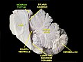
 Deep dissection of brainstem. Lateral view. | |
| Details | |
| Part of | Midbrain |
| Identifiers | |
| Latin | tectum |
| NeuroNames | 465 |
NeuroLex ID | birnlex_1032 |
| TA | A14.1.06.601 |
| TH | H3.11.03.3.01001 |
| TE | E5.14.3.3.1.4.1 |
| FMA | 83902 |
Anatomical terms of neuroanatomy [edit on Wikidata] | |
The tectum (Latin: roof) is a region of the brain, specifically the dorsal (top) part of the midbrain (mesencephalon).[1] The position of the tectum is contrasted with the tegmentum, which refers to the region ventral (lower) to the ventricular system. The tectum is responsible for auditory and visual reflexes.
The optic tectum projects through the reticular formation and interacts with motor neurons in the brainstem.[2] These connections are important for the recognition and reaction to various sized objects which is facilitated by excitatory optic nerve transmitters like L-glutamate.[3] Recent lesion studies have suggested that the tectum has no influence over higher-order motion responses like OMR or OKR[4], but may be more integral to lower-order cues in motion perception like in the identification of small objects.[5]
The tectum is derived in embryonic development from the alar plate of the neural tube.
Contents
1 Structure
2 Function
3 Related terms
4 Additional images
5 See also
6 References
7 External links
Structure
The optic tectum is the visual center in the non mammalian brain which develops from the alar plate of the mesencephalon. It has a laminar organization which allows for different cell types to be present on corresponding layers . One example of the layer specificity is the deep laminae which send output signals away from the tectum toward the motor neurons, specifically the pontine nucleus. The pontine nucleus is located in the basal pons and is responsible for sharing information between the cerebrum and cerebellum (Pontine nucleus). Another example is the superficial laminae which receive input from retinal ganglion cells.
In adult humans, the tectum only consists of the inferior and the superior colliculi.
- The superior colliculus is involved in preliminary visual processing and control of eye movements. In non-mammalian vertebrates it serves as the main visual area of the brain, functionally analogous to the visual areas of the cerebral cortex in mammals.
- The inferior colliculus is involved in auditory processing. It receives input from various brain stem nuclei and projects to the medial geniculate nucleus of the thalamus, which relays auditory information to the primary auditory cortex.
Both colliculi also have descending projections to the paramedian pontine reticular formation and spinal cord, and thus can be involved in responses to stimuli faster than cortical processing would allow. Collectively the colliculi are referred to as the corpora quadrigemina.
The structure is supplied by quadrigeminal artery (a branch of posterior cerebral artery), and superior cerebellar artery.
Function
Disrupting visual experience early on in Zebrafish development results in a change in tectal activity. Changes in tectal activity resulted in an inability to successfully hunt and capture prey.[6] Hypothalamus inhibitory signaling to the deep tectal neuropil is important in tectal processing in zebrafish larvae.[7] The tectal neuropil contains structures including periventricular neurons axons and dendrites. The neuropil also contains GABAergic superficial inhibitory neurons located in stratum opticum.[8] Instead of a large cerebral cortex, Zebrafish have a relatively large tectum that is hypothesized to carry out some of the visual processing that the cortex performs in mammals.[9]
Related terms
The term "tectal plate" (or "quadrigeminal plate") is used to describe the junction of the gray and white matter in the embryo. (ancil-453 at NeuroNames)
Additional images
Tectum

See also
- List of regions in the human brain
- Tectospinal tract
References
^ Bear, Mark F.; Connors, Barry W.; Paradiso, Michael A. (2007). Neuroscience. Lippincott Williams & Wilkins. ISBN 9780781760034..mw-parser-output cite.citation{font-style:inherit}.mw-parser-output .citation q{quotes:"""""""'""'"}.mw-parser-output .citation .cs1-lock-free a{background:url("//upload.wikimedia.org/wikipedia/commons/thumb/6/65/Lock-green.svg/9px-Lock-green.svg.png")no-repeat;background-position:right .1em center}.mw-parser-output .citation .cs1-lock-limited a,.mw-parser-output .citation .cs1-lock-registration a{background:url("//upload.wikimedia.org/wikipedia/commons/thumb/d/d6/Lock-gray-alt-2.svg/9px-Lock-gray-alt-2.svg.png")no-repeat;background-position:right .1em center}.mw-parser-output .citation .cs1-lock-subscription a{background:url("//upload.wikimedia.org/wikipedia/commons/thumb/a/aa/Lock-red-alt-2.svg/9px-Lock-red-alt-2.svg.png")no-repeat;background-position:right .1em center}.mw-parser-output .cs1-subscription,.mw-parser-output .cs1-registration{color:#555}.mw-parser-output .cs1-subscription span,.mw-parser-output .cs1-registration span{border-bottom:1px dotted;cursor:help}.mw-parser-output .cs1-ws-icon a{background:url("//upload.wikimedia.org/wikipedia/commons/thumb/4/4c/Wikisource-logo.svg/12px-Wikisource-logo.svg.png")no-repeat;background-position:right .1em center}.mw-parser-output code.cs1-code{color:inherit;background:inherit;border:inherit;padding:inherit}.mw-parser-output .cs1-hidden-error{display:none;font-size:100%}.mw-parser-output .cs1-visible-error{font-size:100%}.mw-parser-output .cs1-maint{display:none;color:#33aa33;margin-left:0.3em}.mw-parser-output .cs1-subscription,.mw-parser-output .cs1-registration,.mw-parser-output .cs1-format{font-size:95%}.mw-parser-output .cs1-kern-left,.mw-parser-output .cs1-kern-wl-left{padding-left:0.2em}.mw-parser-output .cs1-kern-right,.mw-parser-output .cs1-kern-wl-right{padding-right:0.2em}
^ Precht, W. (1974). "Tectal influences on cat ocular motoneurons". Brain Research. 20 (1): 27–40.
^ Beart, Phillip (1976). "An evaluation of L-glutamate as the transmitter released from optic nerve terminals of the pigeon". Brain Research. 110 (1): 99–114. PMID 6128.
^ Roeser, Tobias (2003). "Visuomotor Behaviors in Larval Zebrafish after GFP-Guided Laser Ablation of the Optic Tectum". Journal of Neuroscience. 23 (9): 3726–3734.
^ Barker, Alison (2015). "Sensorimotor Decision Making in the Zebrafish Tectum". Current Biology. 25 (21): 2804–2814.
^ Avitan, L., Pujic, Z., Mölter, J., Van De Poll, M., Sun, B., Teng, H., Amor, R., Scott, E.K., Goodhill, G.J. (2017) Spontaneous Activity in the Zebrafish Tectum Reorganizes over Development and Is Influenced by Visual Experience. Current biology : CB. 27(16):2407-2419.e4.
^ Heap LA, Vanwalleghem GC, Thompson AW, Favre-Bulle I, Rubinsztein-Dunlop H, Scott EK. Hypothalamic Projections to the Optic Tectum in Larval Zebrafish . Front Neuroanat . 2018;11:135. https://www.frontiersin.org/article/10.3389/fnana.2017.00135.
^ Dunn, Timothy W et al. “Neural Circuits Underlying Visually Evoked Escapes in Larval Zebrafish” Neuron vol. 89,3 (2016): 613-28.
^ Heap LA, Vanwalleghem GC, Thompson AW, Favre-Bulle I, Rubinsztein-Dunlop H, Scott EK. Hypothalamic Projections to the Optic Tectum in Larval Zebrafish . Front Neuroanat . 2018;11:135. https://www.frontiersin.org/article/10.3389/fnana.2017.00135.
External links
| Wikimedia Commons has media related to Midbrain tectum. |
- Diagram
- Photo
"Anatomy diagram: 13048.000-3". Roche Lexicon - illustrated navigator. Elsevier. Archived from the original on 2014-01-01.
Authority control |
|
|---|
This neuroscience article is a stub. You can help Wikipedia by expanding it. |


Comments
Post a Comment